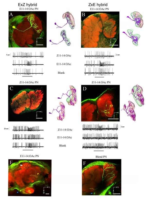
Figure 2.
Dendritic arborization of the projection neurons (PNs) in the macroglomerular complex (MGC) of O. nubilalis F1 hybrids (ExZ, ZxE). Neurobiotin-filled PNs (green) in a α-synapsin-labeled antennal lobe (red). Left panels: confocal stack through a portion of the antennal lobe with part of the neuron visible. Because several confocal sections were overlaid to visualize a large part of the neuron, the otherwise sharp glomerular delineations are somewhat blurred. Right panels: three-dimensional reconstruction of the two large MGC glomeruli (lateral, red; medial, green) and dendritic arborization of the PN (violet). Lower panels: intracellular recording trace of the PN in the upper panel. Stimulation time, 500 ms (grey scale bars). (A) The MGC of a ExZ hybrid male displaying an E11-14:OAc-specific PN with exclusive arborizations in the medial glomerulus. Stimulation dose, 1 ng. (B) The MGC of an ZxE hybrid male displaying an E11-14:OAc-specific PN with exclusive arborizations in the medial glomerulus. Stimulation dose, 10 ng. (C) The MGC of an ExZ hybrid male displaying a Z11-14:OAc-specific PN with exclusive arborizations in the lateral glomerulus. Stimulation dose, 1 ng. (D) The MGC of a ZxE hybrid male displaying a Z11-14:OAc-specific PN with exclusive arborizations in the lateral glomerulus. Stimulation dose, 10 ng. (E) Axonal arborization of the E11-14:OAc-specific PN in the calyces of the mushroom body and in the lateral horn of the protocerebrum. (F) Pheromone blend-specific PN arborization in both large MGC glomeruli, responding only to a 50:50 blend of the Z11-14:OAc and E11-14:OAc. Scale bars in the confocal images, 50 μm.

