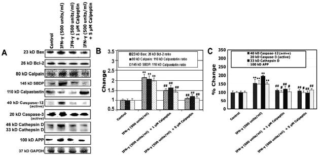
Fig. 5.

Alterations in protein expression of Bax, Bcl-2, calpain, SBDP, calpastatin, caspase-12, and caspase-3, cathepsin D, and APP. Cells were treated with IFN-γ (500 units/ml) for 48 hours. Calpeptin (1 and 5 μM) was added 5 minutes after the IFN-γ addition. (A) Representative Western blots to show levels of 23 kD Bax, 26 kD Bcl-2, 80 kD calpain, 145 kD SBDP, 110 kD calpastatin, active 40 kD caspse-12, active 20 kD caspase-3, 46 kD and 33 kD cathepsin D, 100 kD APP, and 37 kD GAPDH. (B) Determination of changes in Bax:Bcl-2, calpain:calpastatin and SBDP:calpastatin ratios. (C) Determination of percent changes in active 40 kD caspase-12, active 20 kD caspase-3, 33 kD cathepsin D, and 100 kD APP.
