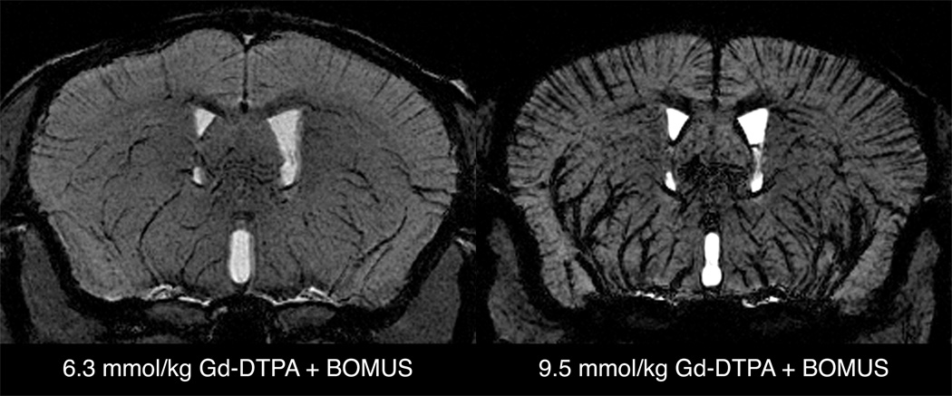
FIG. 10.
Minimum intensity projections of a 600-µm axial slab from SPGR images (high-resolution protocol) from BOMUS-treated animals given high doses of Gd-DTPA. BOMUS allows the Gd-DTPA to enhance the parenchyma of the brain, but high concentration of Gd-DTPA in the bloodstream causes susceptibility-induced loss of signal from the blood and perivascular tissue. This allows the delineation of cortical vessels (running perpendicular to the cortical surface). When the dose of Gd-DTPA is increased to 9.5 mmol/kg, this effect is exaggerated.

