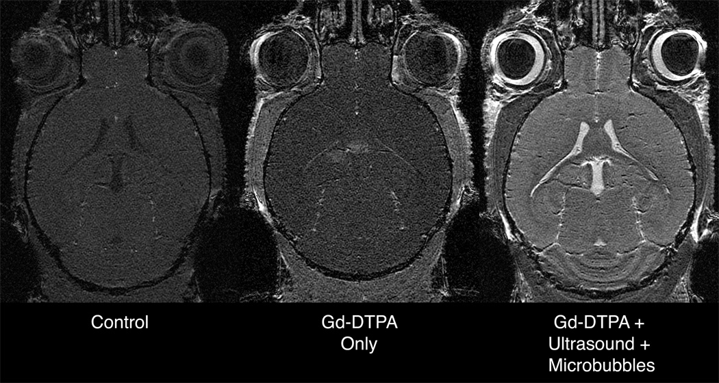
FIG. 9.
High-resolution (52 × 52 × 100 µm3) SPGR images of the mouse brain acquired in vivo in 51 minutes. The control mouse receiving no contrast agent and the mouse receiving only Gd-DPTA have relatively low signal. The animal receiving Gd-DTPA along with BOMUS (microbubbles+ultrasound) shows an increase in SNR of 90% and 63% over the other two. SNR measurements were made in left anterior cortex. For each scan TR = 25ms and FA was adjusted to maximize signal in the anterior cerebral cortex (15, 15, and 25 degrees, respectively).

