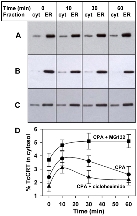
Figure 2. Retrotranslocated TcCRT is degraded by the proteasome.
Cellular fractionation at different time points upon 1 µM CPA addition alone (A), or with a previous incubation for 1 h with (B) 1 mM cycloheximide or (C) 10 µM MG132. (D) Quantification of the fraction of cytosolic fraction of TcCRT shown in gels A–C. Bars represent the standard error of triplicate measurements. In all cases the ER containing fractions were diluted ten fold.

