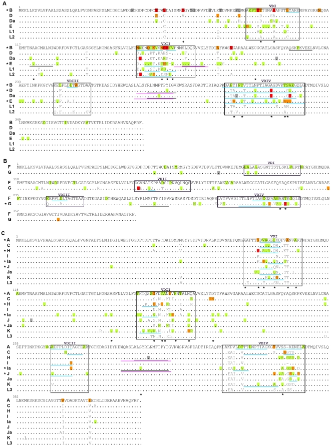
Figure 2. Worldwide mutational pattern of MOMP.
Panels (A), (B), and (C) represent protein alignments of prototype strains according to serogroup B, Intermediate, and C, respectively. Amino acid changes that naturally occur among prototype strains are in grey characters (they do not represent changes among variant specimens and the respective prototype strains). Mutations resulting from the comparison between the 5026 strains isolated in 33 different geographic regions from five continents and the respective prototype strain are highlighted in different colors, corresponding to dissimilar degrees of evolutionary fixation. Green, orange and red represent amino acid changes occurring in strains isolated in a single, 2–4, and ≥5 geographic regions, respectively. Only silent mutations that occurred in strains isolated in ≥5 geographic regions are shown (highlighted in grey). Well-defined B-cell (blue), Th-cell (purple) and CTL (black) antigenic regions are underlined. Degenerated variable sites are marked with an asterisk.

