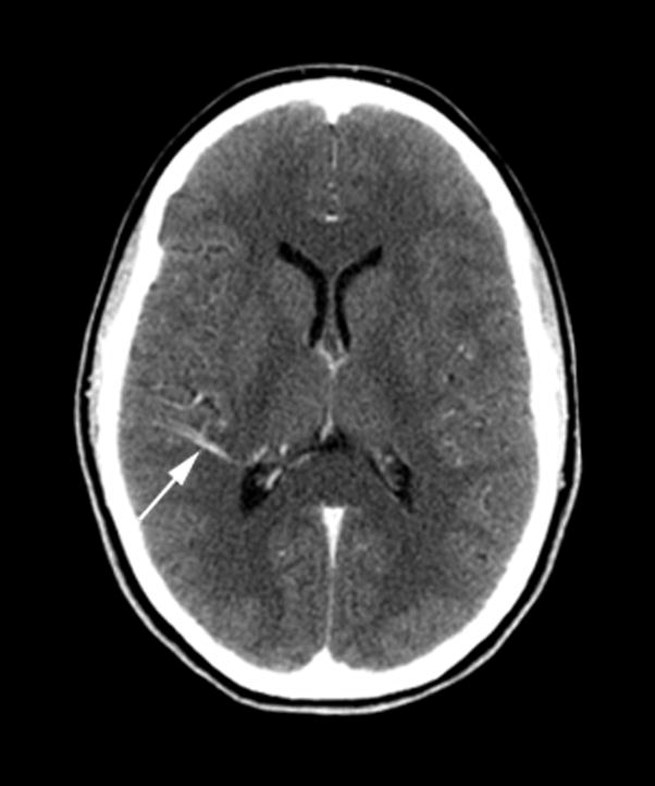
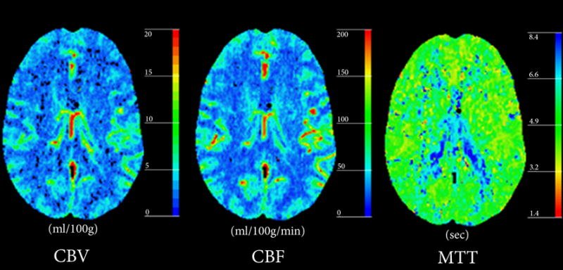
Figure 1. Typical DVA with normal perfusion parameters.


(A) Post-contrast CT shows a right posterior temporal DVA with centripetal drainage (arrow) into choroidal veins in the atrium of the right lateral ventricle.
(B) Perfusion-CT maps do not demonstrate abnormalities in the parenchyma surrounding the DVA.
