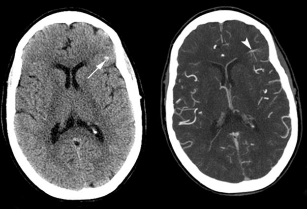
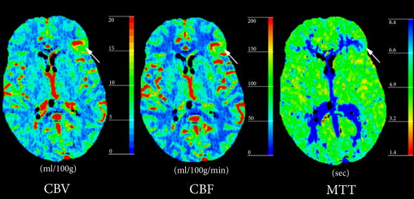
Figure 3. Atypical DVA with abnormal perfusion parameters and associated hemorrhage.

(A) Non-contrast enhanced CT (left) and post-contrast CT (right) show left a frontal lobe DVA within the cortex and subcortical white matter (arrowhead), with a small area of hemorrhage (arrow) in the adjacent parenchyma.
(B) Perfusion CT maps demonstrate regional increase in CBV and CBF surrounding the DVA, and prolonged MTT.

