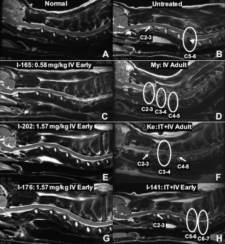
Fig. 1.
Magnetic resonance imaging of the spinal cord. T2-weighted non-contrast sagittal images of the cervical spine. A normal dog (A, age 15 months) showing no abnormalities, and an untreated MPS I dog (B, age 18 months) showing disk protrusion into the canal at C2-3 (arrow) and disk degeneration at C5-6 (circle). Small islands of bony proliferation can be seen within the intervertebral disk at C5-6 (arrowhead). (C, E, G) Spinal MRI of dogs treated with 0.58 mg/kg IV Early (C, age at MRI 17 months) and 1.57 mg/kg IV Early (E, G, age 12 months) showing no abnormalities. (D, F, H) An IV Adult animal at age 29 months (D), an IT+IV Adult dog age 27 months (F) and an IT+IV Early dog age 18 months (H) also show disk degeneration (circles) and disk protrusion (arrows). Circumferential compression is seen in dog Ke (F) at C2-3 (arrow).

