Figure 2.
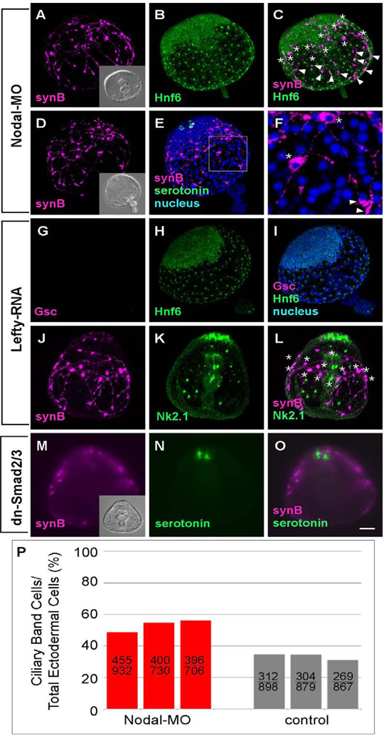
The ciliary band is expanded and shifted to the animal pole in embryos in which Nodal-MO blocks the oral pathway. Ciliary band-associated neurons differentiate, most of the cell bodies are near the Hnf6-expressing ciliary band cells and axons with strongly immunoreactive growth cones project toward the vegetal pole. The position of cell bodies(*) and growth cones (arrowheads) are marked (A–C) Ciliary band cells, identified with anti-Hnf6, cover the lateral ectoderm initially, and become restricted to a thickened epithelium, 10–14 cells wide, surrounding the animal plate. Ectoderm surrounding blastopore thins and expands to eventually cover the vegetal half of the embryo. (B). Serotonergic neurons are present exclusively at the animal plate (D, E). (F) The magnified image of rectangle in (E) shows the neural cell body (*) and growth cones (arrowheads). (G–L) Lefty RNA-injected embryos have the same morphology as embryos injected with Nodal-MO. (G–I) Goosecoid (Gsc) is not expressed (G), indicating that the oral ectoderm has not differentiated. The ectoderm is covered with ciliary band and thin, vegetal ectoderm (H and I). (J–L) Ciliary band-associated neurons are present throughout the entire lateral ectoderm (* mark the position of cell bodies) and the animal plate marker, Nk2.1, is expressed exclusively in the animal plate. (M–O) Embryos injected with RNA encoding a dominant-negative Smad2/3 have the same morphology as embryos injected with Nodal-MO. Ciliary band neurons are spread throughout the entire lateral ectoderm. Serotonergic neurons are present at the animal plate region (N and O). Inset in (M) shows the DIC image of dn-Smad2/3 injected embryo. (P) The number and ratio of ciliary band cells are increased in Nodal morpholino injected embryos. The upper and lower numbers on the column show the counted number of ciliary band cells and total ectodermal cells, respectively. Those cells are counted in three individual embryos of each treatment. Bar = 20µm.

