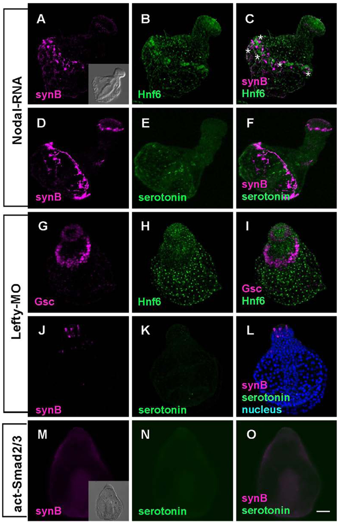
Figure 3.
Embryos are radialized and the ciliary band is reduced to a band 1–2 cells wide that is shifted toward the vegetal pole when Nodal signaling is enhanced. (A–F) A thin Hnf6-positive band contains ciliary band neurons forming tracts of axons that interconnect cells. (C). Serotonergic neurons do not differentiate, because the animal plate is surrounded by oral ectoderm, but non-serotonergic neurons are present in the animal plate (E and F) (* mark the position of cell bodies). (G–L) Lefty-MO-injected embryos are superficially similar to Nodal mRNA-injected embryos, but there is no ciliary band (Hnf6 is restricted to the animal plate) and no ciliary band neurons. (G) Gsc is expressed radially and neither Hnf6-expressing ciliary cells nor synaptotagmin-positive neurons are detected in the lateral ectoderm (H–L). (M–O) Constitutively activated Smad2/3 mRNA-injected embryos have a similar form to other embryos with enhanced Nodal signaling, but they are like the Lefty-MO- injected embryos in lacking ciliary band neurons. Inset in (M) shows the DIC image of act-Smad2/3 injected embryo. Bar = 20 µm.

