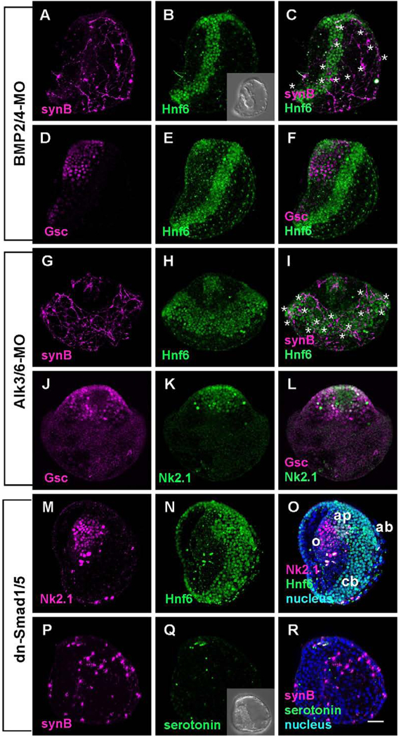
Figure 4.
The embryos in which BMP2/4 signaling is blocked have wider than normal Hnf6-expressing ciliary bands that are shifted vegetally and unpatterned neurons. (A) Ciliary band neurons differentiate in association with the ciliary band and the aboral ectoderm. (B) The ciliary band is wider (5–6 cells in width) and shifted toward the aboral ectoderm. (C) Merged image of (A) and (B) (* mark the position of cell bodies). (D) Oral ectoderm identified with anti-Gsc surrounds the animal plate. (E) Hnf6 marks the animal plate and shifted ciliary band away from animal plate. (F) Merged image of (D) and (E). (G–L) Alk3/6 is the only BMP receptor in the sea urchin genome, yet morpholinos produce embryos with a more severe phenotype with a greatly expanded, radialized ciliary band (* mark the position of cell bodies). (G) Neurons differentiate in association with the ciliary band and the aboral ectoderm. (H) Hnf6 reveals that the ciliary band is shifted vegetally, does not connect with the animal plate, and is 10–12 cells wide. (J–L) An oral ectoderm, expressing Gsc surrounds the animal plate (Nk2.1). (M–R) Embryos in which a dominant-negative form of Smad1/5 is injected are similar to embryos in which Alk3/6 receptor expression is suppressed. (M–O) The animal plate, identified with an Nk2.1 antibody, is surrounded by oral ectoderm and there is a thickened ciliary band (10–12 cells wide; animal pole view). (P–R) Ciliary band neurons differentiate in association with the ciliary band and project neurites with strongly immunoreactive growth cones toward the vegetal end of the embryo. Inset in (Q) shows the DIC image of dn-Smad1/5 injected embryo. Bar = 20µm; o; oral ectoderm; ap, animal plate; cb, ciliary band; ab, aboral ectoderm.

