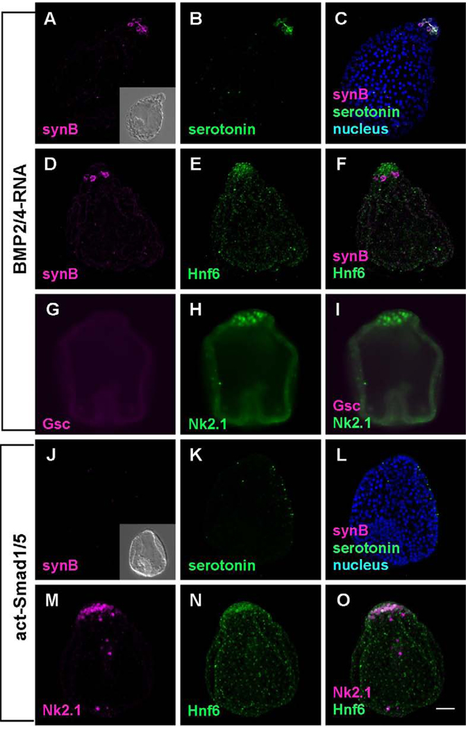
Figure 5.
The embryos in which BMP2/4 signaling is enhanced are radialized and lack ciliary bands and ciliary band neurons. (A–I) BMP2/4- misexpressing embryos. (A–F) The embryos lack synaptotagminB-expressing neurons and Hnf6-expressing ciliary band cells throughout the lateral ectoderm. However, serotonergic neurons are invariably present in the animal plate (B and C), indicating the animal plate is not influenced by the misexpression of BMP2/4. (G–I) Embryos lack oral ectoderm expressing Gsc, but express NK2.1 in the animal plate. (J–O) The embryo in which constitutively activated Smad1/5 mRNA has been injected is similar to BMP2/4-misexpressing embryos. They have an animal plate identified with Hnf6 and Nk2.1 (M–O), but no serotonergic neurons differentiate because of the intracellular activation of Smad1/5 (K–L). Inset in (J) shows the DIC image of dn-Smad1/5 injected embryo. Bar = 20µm.

