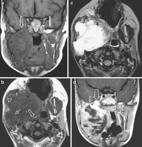
Fig. 1.
Masticator space eRMS in a 4-year-old boy presenting with a rapidly progressive right side preauricular mass. a Coronal SE T1-W image without IV contrast demonstrates obliteration of the fat planes (arrow) of the MS and the PPS and of the sphenoid wing on the right side [compare to the normal left side (white arrow)]. Also note abnormal configuration and signal of the right mandible, better shown on the axial SE T1-W images (b). b Axial SE T1-W image shows tumour bulging through the mandible underneath the masseter muscle with preservation of the fat plane (arrowhead). The PPS is compressed and the fat is displaced posteriorly and medially (arrow), indicating that the origin of the tumour is not in the PPS but in the MS. Extension of tumour through the stylomandibular foramen into the parotid gland (small arrow). c The mass is of high signal intensity throughout on axial SE T2-W sequence. d Coronal SET1 image after i.v. contrast: heterogeneous enhancement with many necrotic areas. Tumour extention through the foramen ovale into the right cavernous sinus (large arrow). Note also subtle dural enhancement (small arrow), which was interpreted as tumour extension, but might have been reactive changes only, according to thickness < 5mm

