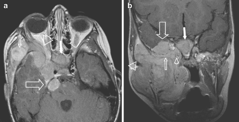
Fig. 2.
Four-year-old girl, presenting with a right side preauricular mass and cranial nerve palsy (ptosis). A huge eRMS originating within the MS was found with involvement of the greater wing of the sphenoid bone and orbital apex on the right side, with massive perineural extention along the 2nd and 3rd branches of the right trigeminal nerve and involvement of the cisternal portion. a Axial contrast-enhanced SE T1-W image shows a mass within the masticator space (small arrow), abnormal enhancement of the sphenoid wing (arrowhead), the clivus (white arrow) and multiple masses along the 2nd branch of the right trigeminal nerve (pterygopalatine fossa, foramen rotundum, cavernous sinus) extending into the posterior fossa with involvement of the cisternal portion of the trigeminal nerve (large arrow). b Coronal contrast-enhanced SE T1-W image of the same patient, shows involvement of the right MS, along the temporalis muscle (large arrowhead), into the masseter muscle. Tumour extends into the foramen rotundum (small arrowhead), the sphenoid wing (small arrow) with massive dural invasion (>5 mm) (large arrow) and invasion of the clivus (white arrow)

