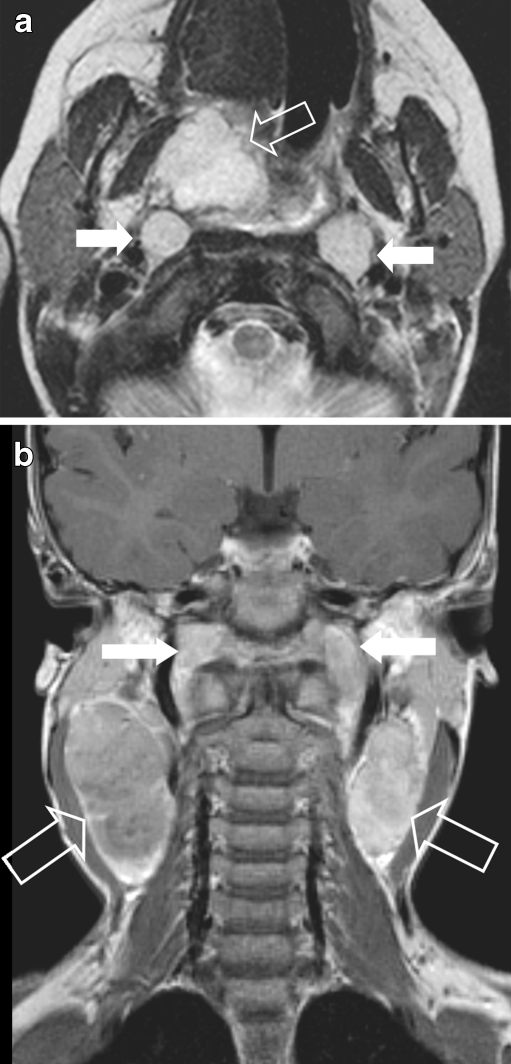
Fig. 9.
Nasopharyngeal e-RMS in a 4-year old girl, presenting with massive cervical lymphadenopathy, suggesting nasopharyngeal cancer. a Axial SET2 weighted image showing a high signal lesion (arrow) within the right naso - oropharynx and enlarged retropharyngeal lymph nodes (white arrow). b Coronal SET2 image showing massive, bilateral cervical lymphadenopathy (open arrows & white arrows). Final histology: e-RMS

