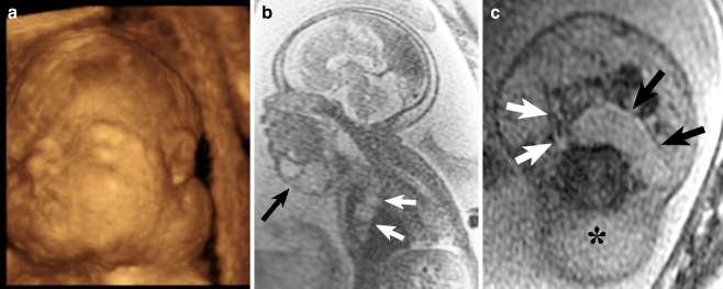
Fig. 6.
Lymphatic malformation. a 3-D sonographic image in a 25-week fetus with suspected neck mass on prior US at an outside facility demonstrates a mass arising from the left lateral aspect of the fetal neck. b Sagittal SSFSE T2-W image in the same patient as Fig. 7 obtained at 31 weeks demonstrates a multiseptated T2 hyperintense mass in the anterior neck (black arrow) with extension into the anterior mediastinum (white arrows), most consistent with lymphatic malformation. Secondary signs of airway obstruction were not present. c Axial SSFSE T2-W image demonstrates a cystic retropharyngeal component of the multiseptated mass (black arrows) that displaces the oropharynx (white arrows) anterolaterally. Hydrops of the soft tissues posterior to the cervical spine (asterisk) is also seen. This baby died shortly after birth

