
Figure 2.
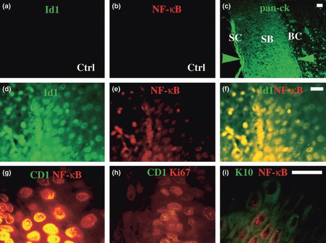
Id1, NF‐κB, cyclin D1 and Ki67 are highly expressed in cholesteatoma tissues. Normal middle ear epithelium was negative for Id1 (a) and activated NF‐κB (b), whereas cholesteatoma produced abundant pan‐cytokeratins (pan‐ck) in the basal cell (BC), and suprabasal (SB) and stratum corneum (SC) layers (c, between green arrowheads). Id1 (d) and activated NF‐κB (e) were co‐expressed in the basal cell (BC) layer (f) of cholesteatoma tissues. Similarly, activated NF‐κB and cyclin D1 (g) as well as cyclin D1 and Ki67 (h) were co‐expressed in the basal layer; while keratin 10 and NF‐κB (i) were co‐expressed in the suprabasal layer of cholesteatoma epithelium. Bar = 10 μm applying to the same row; Ctrl, control middle ear tissue (a and b); and cholesteatoma tissue (c–i).