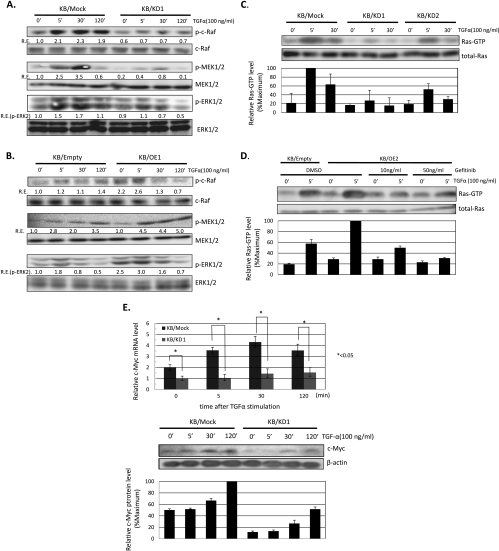
Figure 3.
CIN85 preferentially promotes activation of the Ras-ERK pathway critical for the growth of HNSCCs. (A and B) Western blot assays for phospho-c-Raf, -MEK1/2, and -ERK1/2 in KB/mock, KB/KD1, KB/empty, and KB/OE1 cells. Cells were treated with 100 ng/ml of TGF-α for the indicated times, and cell lysates were prepared. Relative expression (R.E.) was calculated as described in Figure 2. The results are representative of three independent experiments. (C) Effect of CIN85 expression on Ras activation in KB/mock and KB/KD cells. Cells were treated with 100 ng/ml of TGF-α for the indicated times, and the abundance of the activated forms of Ras (Ras-GTP) and total cellular Ras proteins was determined. The levels of specific bands were quantified, and those of Ras-GTP were normalized with respect to those of corresponding Ras. The resulting values were expressed as the percentage in reference to that of KB/mock stimulated with TGF-α for 5 minutes. The results are representative of two independent experiments. (D) Gefitinib blocks CIN85-induced enhancement of Ras activation. Cells were treated with the indicated concentrations of gefitinib for 30 minutes before TGF-α stimulation, and the abundance of activated forms of Ras (Ras-GTP) and total cellular Ras proteins was determined. The resulting values were expressed as the percentage in reference to that of KB/OE2 stimulated with TGF-α for 5 minutes. The results are representative of two independent experiments. (E) Effect of CIN85 expression on c-Myc expression in KB/mock and KB/KD cells. Cells were treated with 100 ng/ml of TGF-α for the indicated times, and total RNA and proteins were extracted. Relative expression of c-Myc mRNA was determined by quantitative RT-PCR assays (upper panel). The values are the mean ± SD of three independent experiments. *P < .05. The levels of c-Myc protein were detected by Western blot assays (lower panel). β-Actin was served as a loading control. The resulting values were expressed as the percentage in reference to that of KB/mock stimulated with TGF-α for 120 minutes.

