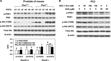
Figure 5.
SDC-1 and n-3 PUFA reduce PDK-1 and Akt phosphorylation in vivo and in vitro. (A) Protein extracts from the prostates of 8-week-old PtenP+/+ and PtenP-/- mice fed n-3 PUFA- and n-6 PUFA-enriched diets were used for Western blot analysis of p-PDK1(S243), PDK1, p-Akt (S473 and T308), total Akt, and β-actin. The mean ± SD for ratios of P-PDK1 to PDK1 and p-Akt (S473 and T308) to Akt are shown in the bottom graph. (B) PC3 cells were treated with SDC-1 ectodomain (50, 100, and 150 nM) and DHA-BSA (30 and 60 µM) for 48 hours. Protein extracts were used for Western blot analysis of p-PDK1, PDK1, p-Akt (S473 and T308), total Akt, and β-actin.

