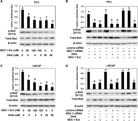
Figure 7.
DHA inhibits Bad phosphorylation through SDC-1. PC3 (A) and LNCaP (C) cells were treated with DHA (30 and 60 µM) or SDC-1 ectodomain (50, 100, and 150 nM) for 48 hours. Total proteins were used to measure p-Bad (S112) and total Bad by Western blot assay. PC3 cells (B) and LNCaP cells (D) were transfected with control siRNA or SDC-1 siRNA for 6 hours and then supplemented with growth medium containing 1% FBS for 24 hours. Cells were treated with DHA (60 µM) or SDC-1 ectodomain (150 nM) for 48 hours before harvest. Total Bad and p-Bad (S112) were measured by Western blot assay. All data shown are representative of three independent experiments. Relative ratios and SEMare shown in graphs, and values representing the mean ± SD (n = 3) with different letters are significantly different (P < .05).

