FIG. 3.
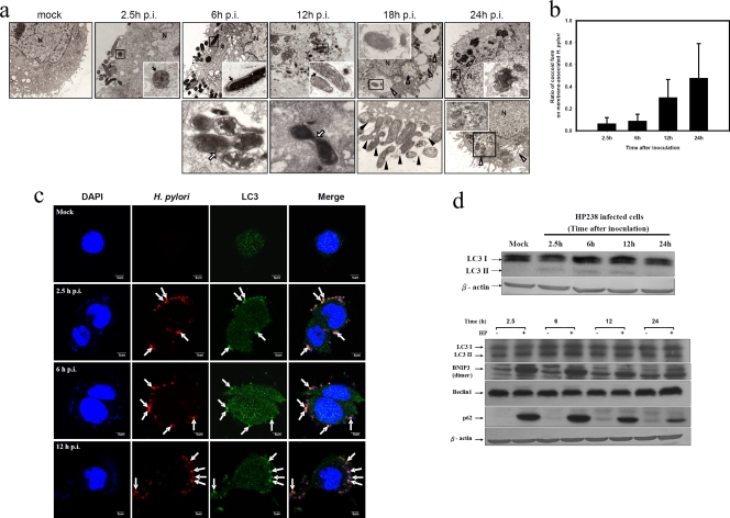
Autophagy induction by H. pylori in AGS cells. (a) H. pylori resides and replicates in double-layer vesicles. AGS cells were infected with HP238 at an MOI of 50 for 1 h. The infected cells were collected, fixed, and observed with electron microscopy at various time points postinfection. The fate of H. pylori postinfection is shown in a time sequence, as indicated on the figure. N, nucleus; MV, multiple-layered vesicle. Black arrows indicate double-layer membranes, whereas white arrows indicate the replicating H. pylori bacteria. White arrowheads indicate the multiple vacuoles, whereas black arrowheads indicate the coccoid forms of H. pylori. (b) The ratio of spiral to coccoid forms on the infected cells at various time points postinfection is shown. (c) LC3 punctate formation colocalized with H. pylori. The arrows indicate the colocalization of LC3 and H. pylori-containing vesicles. (d) Autophagic flux was induced post-H. pylori infection. LC3-II conversion, BNIP3, and p62 were induced postinfection. Each experiment was repeated at least two times with the same result.

