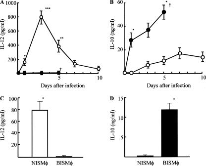
FIG. 3.
Detection of IL-12 and IL-10 in homogenates of MRSA infection site tissues and production of IL-12 and IL-10 by Mφ isolated from MRSA infection site tissues. A square (15 by 15 mm) of infection site tissue was excised from normal mice (n = 6; open circles) or burned mice (n = 6; filled circles) 1, 3, 5, 7, and 10 days after the MRSA infection (108 CFU/mouse). Tissues were homogenized, and supernatants obtained were assayed for IL-12 (A) and IL-10 (B) by ELISA. Data are means ± SEM of results from three different experiments. *, P < 0.05; **, P < 0.01; ***, P < 0.001; †, dead. Three days after intradermal infection of MRSA (108 CFU/mouse), Mφ were isolated from the infection site tissues of normal and burned mice (3 mice each). Then, cells obtained were adjusted to 5 × 10⁵ cells/ml and cultured for 48 h. Culture fluids obtained were tested for IL-12 (C) and IL-10 (D) by use of ELISA. Data are means ± SEM of results from three different experiments. *, P < 0.001.

