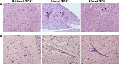
FIG. 3.
PbA-infected PKCθ−/− mice display reduced pathological changes in the brain. Brains were removed from control uninfected or PbA-infected PKCθ+/+ and PKCθ−/− mice 7 days postinfection, fixed in paraformaldehyde, and embedded in paraffin, and 5-μm sections were stained with hematoxylin and eosin. (A) Representative sections of the cortex of a control PKCθ+/+ mouse and PbA-infected PKCθ+/+ and PKCθ−/− mice, demonstrating hemorrhagic foci (arrows) in the brain cortex (magnification, ×100). (B) Increased accumulation of erythrocytes and leukocytes in cerebral vessels of PKCθ+/+ mice (magnification, ×200). The results are representative of two independent experiments, with four mice analyzed per group.

