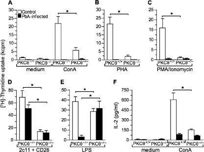
FIG. 6.
In vitro proliferation and IL-2 production by spleen cells from control and PbA-infected PKCθ+/+ and PKCθ−/− mice in response to mitogenic stimuli. Spleen cells from control or PbA-infected PKCθ+/+ and PKCθ−/− mice (day 12 p.i.) were cultured in wells of microtiter plates (4 × 10⁵ cells/200 μl/well) in triplicates in the presence of medium only or 2.5 μg of ConA/ml (A), 10 μg of PHA/ml (B), 100 ng of PMA/ml plus 200 ng of Ionomycin/ml (C), anti-CD3 (ascites, 145-2C11 [dilution, 1:500]) plus anti-CD28 (ascites [dilution, 1:250]) (D), or 10 μg of LPS/ml (E). After 3 days in culture, cells were pulsed with 1 μCi of [methyl-3H]thymidine/well for 6 h and then harvested, and [3H]thymidine incorporation was determined for each group. The data are presented in a bar graph as means ± the SD. (G) IL-2 levels in control and ConA-stimulated cell culture supernatants were determined by ELISA. The data are presented as means ± the the SD of four mice per group and are representative of two independent experiments. *, P < 0.05 (PKCθ+/+ versus PKCθ−/−) as determined by Student t test.

