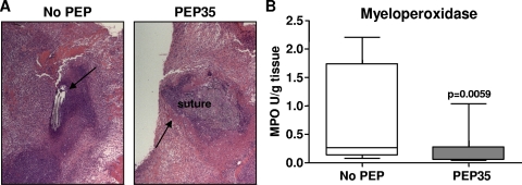
FIG. 2.
PEP35 does not result in sustained neutrophil influx at the S. aureus wound infection site. (A) Surgical wounds were established in WT mice and infected with S. aureus strain PS80 (102 CFU) alone or in combination with PEP35 (100 μg) or a control peptide. Wound tissue was excised on day 5 after the induction of infection, formalin fixed, and embedded in paraffin. Tissue sections were H&E stained to visualize inflammatory infiltration around the wound suture site (representative sections of n = 5 individual mice per group). (B) Wound tissue was also excised on day 5 and homogenized, and the tissue MPO levels were quantified (n = 12 to 14 mice per group). The median is represented by the horizontal bar within the box. The upper and lower boundaries represent the 25th to 75th percentiles of the data, and whiskers represent the 10th and 90th percentiles of the data.

