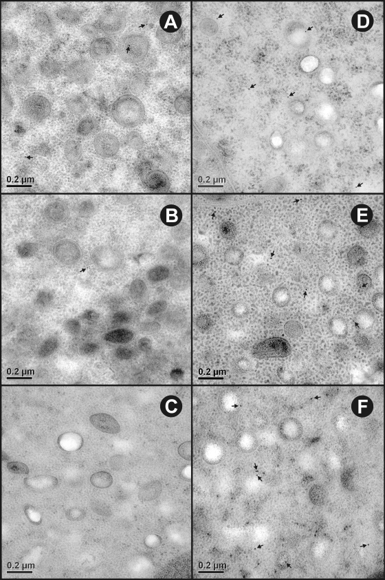
FIG. 4.
Ultrastructural immunolocalization of To16, To18, and To45W in the penetration gland type 2 (PG2) cell of T. ovis nonactivated oncospheres. (A to C) Sections probed with control antisera raised against the MBP fusion protein had a negligible amount of nonspecific staining. (D to F) Sections probed with specific antisera raised against To16, To18, and To45W, respectively, revealed immunogold particles (arrows) distributed sparsely in the cytoplasm and secretory granules of the PG2 cell.

