FIG. 5.
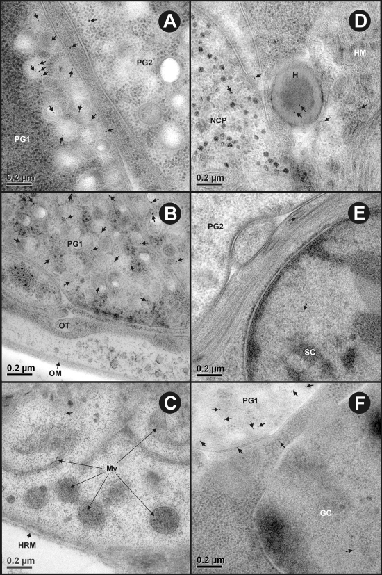
Representative images showing immunolabeling density in various ultrastructural features and cells of T. ovis nonactivated oncospheres reacted with antisera against To18. (A) Oncospheral penetration gland cell types 1 (PG1) and 2 (PG2) showing the difference in their labeling density (arrows). (B) Immunogold particles (arrows) are restricted to the PG1 cell, showing no reactivity with the oncospheral membrane (OM) or tegument (OT). (C) The somatophoric pole of the oncosphere showing no labeling on the hook region membrane (HRM) and microvilli (Mv) of the oncospheral tegument (arrows). (D to F) A low background level of labeling (arrows) in Hook (H), hook muscles (HM), nerve cell process (NCP), somatic (SC), and germinative (GC) cells.

