FIG. 3.
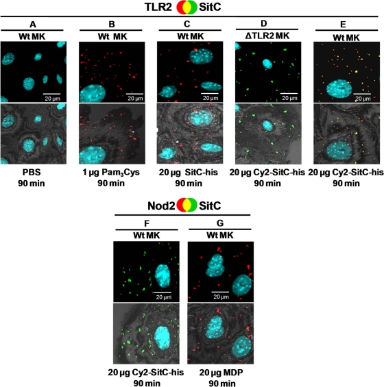
Cy2-SitC-His is incorporated into MKs and colocalizes with TLR2. Confocal images show primary MKs stained intracellularly with a TLR2 or Nod2 antibody from rabbits (detected by a Cy3-conjugated anti-rabbit antibody [red]). Nuclei were stained with DAPI (blue). SitC-His was labeled with Cy2 (Cy2-SitC-his; green). The upper panels show merged images; colocalization events are visualized in yellow. The lower images show an overlay of the fluorescence merge and the host cell, acquired in reflection mode of the confocal microscope. (A) PBS control. (B and C) TLR2 accumulated after stimulation with Pam3Cys (B) and with nonlabeled SitC-His (C). (D) Cy2-SitC-His was incorporated into TLR2-deficient MKs. No TLR2 was detectable in those cells. (E) TLR2 accumulated and colocalized with Cy2-SitC-His in wild-type MKs. (F) Internalized Cy2-SitC-His did not lead to accumulation of Nod2. (G) Stimulation with muramyl dipeptide (MDP) led to an accumulation of Nod2. Images of cells shown are representative of the cells observed in each dish and are representative of three experiments.

