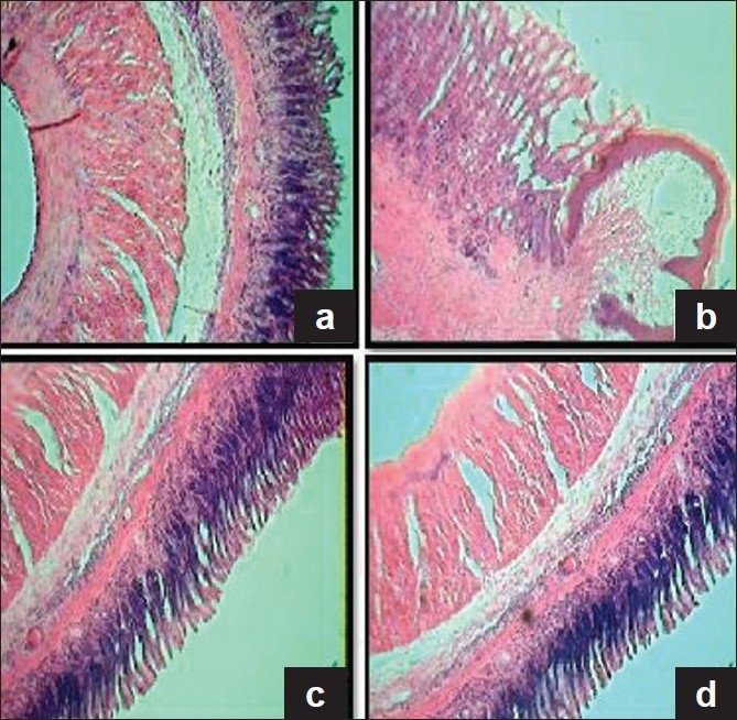
Figure 5.

Histologic examination of gastric mucosal section in the control and experimental rats (H&E, 4×). (a) Section of gastric mucosa obtained from control rat, showing intact cellular architecture. (b) Section of gastric mucosa obtained from AA induced rat, showing marked ulcer crater and damage of submucosal layer, and muscularis region has been replaced by necrotic tissue. (c) Section of gastric mucosa obtained from CQE and AA administered rat, showing absence of ulcer crater, restitution of submucosal layers along with normal glands, complete clearing of inflammatory exudates and reepithelization. (d) CQE alone treated rat depicting normal appearance in the arrangement of mucosal layers almost similar to that of control rat
