Fig. 5.
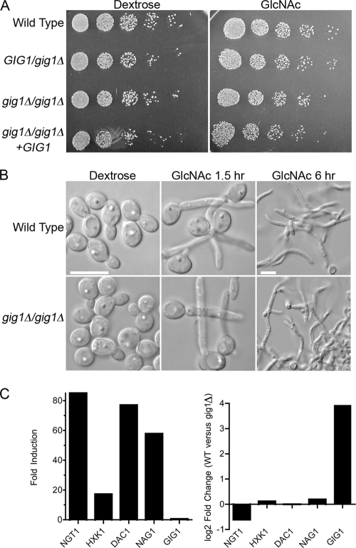
Growth and morphogenesis of gig1Δ mutant cells. (A) A 10-fold dilution series of the indicated C. albicans strains was spotted onto synthetic medium agar plates containing either glucose or GlcNAc at 25 mM. The cells were grown for 2 days at 30°C and were then photographed. The strains used included the wild-type control DIC185, YJA22 (GIG1/gig1Δ), YJA25 (gig1Δ/gig1Δ), and YJA26 (gig1Δ/gig1Δ + GIG1). (B) Cells were grown overnight in synthetic dextrose medium, washed, resuspended in synthetic medium containing either dextrose or GlcNAc at 50 mM, and then grown at 37°C for the indicated times. The GlcNAc 6-h samples are shown at a lower magnification to allow the visualization of long hyphal structures. Bars, 10 μm. (C) (Left) Fold induction of the indicated genes after a 2-h incubation of the gig1Δ strain in GlcNAc, as determined by a microarray experiment. (Right) Direct comparison on the same microarray of wild-type control cells and gig1Δ cells that were shifted to GlcNAc for 2 h.

