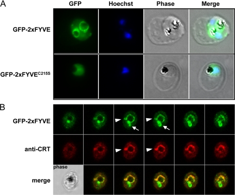
Fig. 4.
GFP fluorescence in parasites expressing GFP-2×FYVE or GFP-2×FYVEC215S. (A) Cells were fixed and analyzed for GFP fluorescence. Nuclei were stained with Hoechst stain (blue stain in the merged images). (B) Colocalization of GFP-2×FYVE and the food vacuole membrane marker CRT using rabbit anti-CRT. Serial images of a Z-stack acquisition (0.38 μm step) are displayed. Arrowheads indicate protrusions from the food vacuole membrane that are colabeled with CRT and GFP-2×FYVE, while arrows indicate zones of intense GFP-2×FYVE staining in the absence of the food vacuole marker. A corresponding phase-contrast image is shown in the lower left corner.

