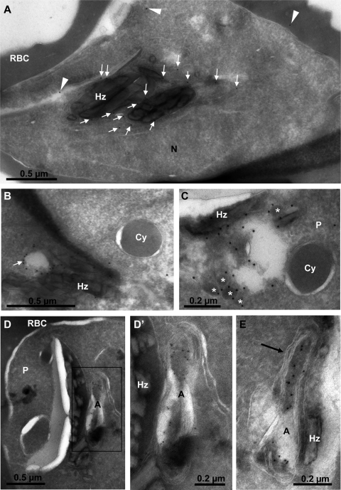
Fig. 6.
Localization of GFP-2×FYVE by cryoimmunoelectron microscopy. (A) Section through a young schizont showing most of the gold label on or near the food vacuole membrane (arrows). Arrowheads point to what should be considered the background. Hz, hemozoin crystals; N, parasite nucleus; RBC, erythrocyte cytosol. (B and C) The membranes of electron-lucent (B) (arrow) and electron-dense (C) (asterisks) vesicles neighboring the food vacuole (Hz) are strongly labeled. Cytostomal vesicles (Cy) are not labeled. (D to E) General (D) and close-up (D′ and E) views of young schizonts showing the strong labeling of the apicoplast, mostly within the lumen. The four membranes of the apicoplast are clearly identified (E, arrow). A, apicoplast; P, parasite.

