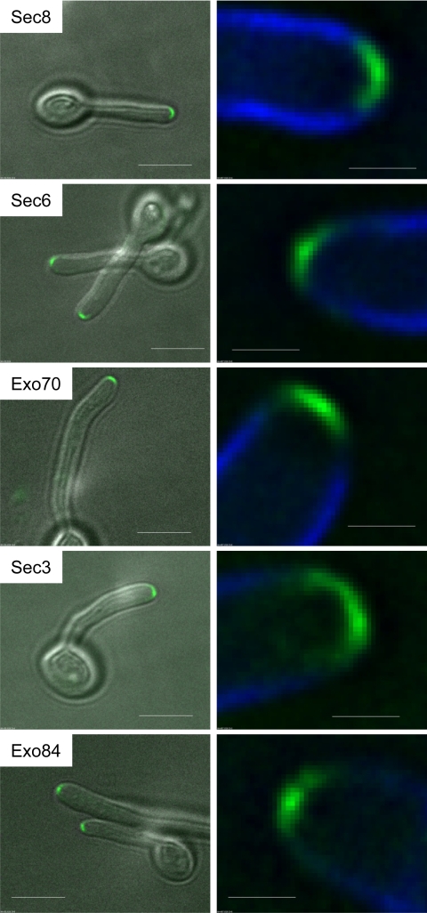
Fig. 1.
Exocyst components localize to a surface crescent. Hyphae expressing the indicated fusions to YFP were imaged 90 min after stationary-phase yeast cells were induced to form hyphae. Images are projections of the deconvolved Z-stack. All images are representative. Scale bars: left panels, 5 μm; right panels, 1 μm.

