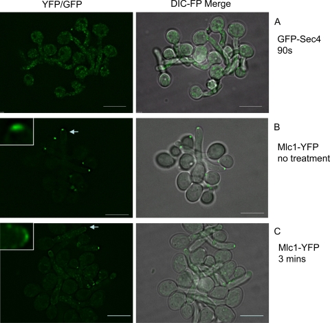
Fig. 5.
Spitzenkörper components depolarize rapidly upon disruption of the actin cables with cytochalasin A. Hyphae of the indicated strains were treated with 10 μM cytochalasin A and images recorded after the indicated times. The strain expressing Mlc1-YFP is shown pretreatment, as its localization is not shown elsewhere in this paper. In all hyphae it localizes to a bright subapical spot (e.g., arrowed tip, enlarged in the inset). After 3 min of treatment with cytochalasin A, these have largely disappeared. In some hyphae Mlc1-YFP remains in a fainter surface crescent (e.g., arrowed tip, enlarged in the inset). The experiment was initiated at 60 min after inoculation of stationary-phase yeast cultures into synthetic complete medium plus 20% serum and incubation at 37°C. Scale bars, 5 μm.

