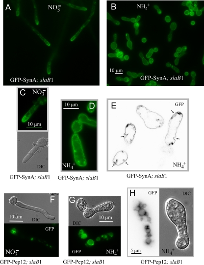
Fig. 4.
Abolishment of SynA endocytic recycling by SlaB downregulation. (A) slaB1 hyphae on nitrate, showing normal, polarized plasma membrane localization of SynA. (B) A general view of slaB1 cells after overnight incubation on ammonium at 25°C. (C) slaB1 germling cultured on nitrate for 9 h, showing that SynA polarization is already conspicuous at this early stage of polarity maintenance. (D) slaB1 cell cultured overnight on ammonium. SynA is not polarized, and the SynA-GFP-labeled plasma membrane is ruffled. Panels C and D are at the same magnification. (E) Confocal image of SynA-GFP (inverted contrast) showing the large cavities that are formed when slaB1 cells are cultured on ammonium. This plane is a frame of Movie S3 in the supplemental material, which should be consulted to fully appreciate the size of these cavities. (F) slaB1 germling expressing GFP-Pep12, cultured on nitrate. GFP-Pep12 localizes to the vacuolar membrane and endosomes. (G) slaB1 germling expressing GFP-Pep12, cultured on ammonium. GFP-Pep12 localizes to the vacuolar membrane as on nitrate, but the abnormal cells show enlarged vacuoles. (H) Another example of a slaB1 germling expressing GFP-Pep12 that had been cultured on ammonium. It shows extensive vacuolization.

