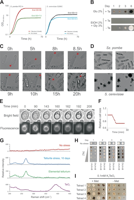
Fig. 1.
Tellurium is accumulated in yeast following Te(IV) exposure. (A) Growth curves were recorded for S. cerevisiae and S. pombe cells microcultivated with and without 0.1 mM K2TeO3 for 70 h by automated measurements of medium OD/turbidity. (B) S. cerevisiae cells (BY4741) were spotted onto fermentative (glucose [Glu]) and respiratory media with and without 0.5 mM K2TeO3 and cultivated for 6 days. EtOH, ethanol; Gly, glycerol. (C) Cellular darkening and plaque formation in a single S. pombe cell (red arrow) through 20 h of 0.5 mM K2TeO3 stress. (D) Cellular darkening and plaque formation in S. pombe (3 days, liquid cultivation) and S. cerevisiae (11 days, solid medium cultivation) after exposure to 0.5 mM K2TeO3. (E) Tellurium plaque formation with respect to the vacuole was followed by time-lapse microscopy of S. cerevisiae S288c cells stained with a fluorescent vacuolar membrane dye. The time sequence shows a typical cell. Time zero is ∼19 h after the addition of 0.5 mM K2TeO3. See also videos S1 to S3 in the supplemental material. (F) The relative cell size decreases to about 75% of the initial cell size in Te(IV)-exposed cells following collapse of the vacuole. The relative size of a typical S. cerevisiae cell is shown. Time zero is ∼21 h after the addition of 0.5 mM K2TeO3. (G) Raman spectra of S. cerevisiae cells grown for 11 days in the absence or presence of 2 mM K2TeO3, as well as Raman spectra of oxidized (TeO2) and nonoxidized elemental tellurium, Te. (H) Tellurium accumulation and Te(IV) tolerance of WT S. cerevisiae strains BY4741 (MATa met17Δ0) and BY4742 (MATα lys2Δ0), which are isogenic except for their auxotrophic markers and mating types. (I) High tellurium accumulation and Te(IV) sensitivity cosegregate with the met17Δ0 marker in the F1 haploid progeny of the BY4741 × BY4742 cross. Shown is colony growth 2 days after replica plating from rich medium onto a medium supplemented with 0.1 mM K2TeO3. Met, methionine.

