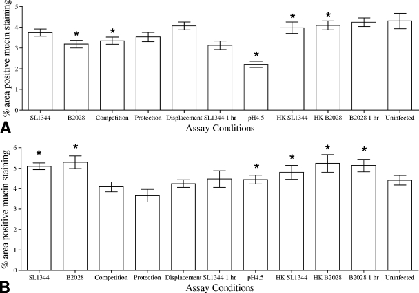
FIG. 4.
Analysis of the percentages of areas of jejunal IVOC tissues staining positive for AB (A) or PAS (B) following in vitro competitive exclusion assays using L. plantarum JC1 (B2028). Uninfected tissue, heat-killed L. plantarum and S. Typhimurium, and assay medium at pH 4.5 were used as control conditions for both tissues. Results are presented as the means ± standard deviations of the means. Asterisks indicate conditions where the percentages of areas of the IVOC tissue exhibiting positive mucin staining were significantly (P < 0.05) different from the uninfected control results.

