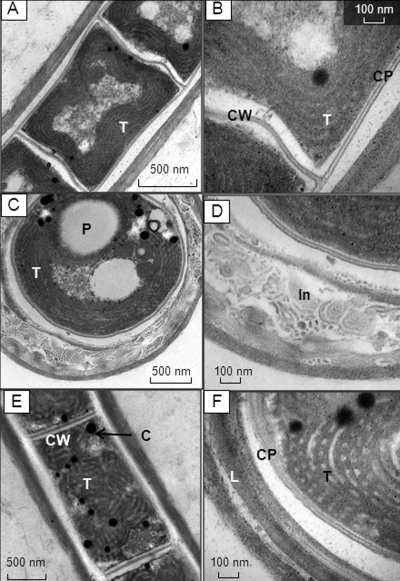
FIG. 2.
TEM micrographs of JSC-1 cells grown with 40 μM FeCl3·6H2O. (A to D) Sections of isodiametric cells; (E and F) sections of elongated cells; (A, B, and E) longitudinal sections; (C, D, and F) cross sections. T, thylakoids; CP, cytoplasmic membrane; CW, cross wall; P, polyphosphate body; In, inclusions within external sheath; C, cyanophycin granules.

