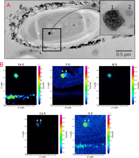
FIG. 6.
TEM views of extracellular and intracellular iron-rich particles found in JSC-1 cells incubated with 600 μM FeCl3·6H2O and 0.04 mM Na2HPO4·7H2O. These cells were not stained with Os. (A) TEM view of a JSC-1 cell encrusted with external Fe-bearing precipitates and containing an electron-dense, internal, Fe-rich particle ∼100 nm in size (insert, image numbered 1) and a satellite body ∼30 nm in size (insert, image numbered 2). (B) Quantitative element maps for internal and external particles show distribution patterns for Fe, P, Al, Ca, and O. The electron dense particle 1 contains major amounts of Fe, P, and O with minor amounts of Al and Ca. Particle 2 contains P, Ca, and O, with no detectable Fe (color panels).

