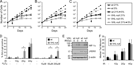
FIG. 2.
VHL loss sensitizes cells to oxidative stress. (A to C) Growth assays of wild-type and VHL-null MEFs grown at 21% oxygen or 5% oxygen or grown for 4 days (A), 8 days (B), or 12 days (C) at 21% oxygen before being moved to 5% oxygen. The arrows indicate when the cells were transferred to 5% oxygen. (D) Quantification of SA-β-Gal staining of cells grown at 5% O2 12 days after treatment with various doses of ionizing radiation (*, P < 0.001), or treated with H2O2 and assayed after 8 days. (E) Western blot analysis of wild-type and VHL-null cells before and 30 min after 4 Gy of IR for γH2AX staining. (F) Quantification of the median tail moments of nuclei in denaturing comet assays with or without IR treatment, as indicated. The error bars represent standard deviations.

