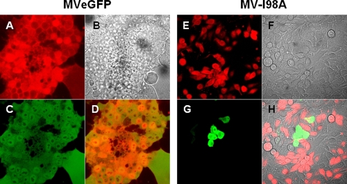
FIG. 2.
Membrane fusion after infection with wild-type or mutant viruses. Adherent Mel624 cells were infected with MVeGFP or MV-I98A and overlaid on target uninfected cells labeled with cell tracker orange. The presence/absence of fusion was detected as previously discussed and the Spearman's correlation coefficient calculated. The images are as follows: A and E, target cells labeled orange with the red filter only; B and F, phase contrast images; C and G, green filter showing infected cells expressing GFP; D and H, merged images showing colocalization (or lack) of signals. Fusion was apparent in MVeGFP-infected cells, as giant multicell syncytia were present (D) (Spearman's ρ = 0.52); however, these syncytia were absent from cells infected with MV-I98A (H), as no syncytia were observable (Spearman's ρ = −0.28).

