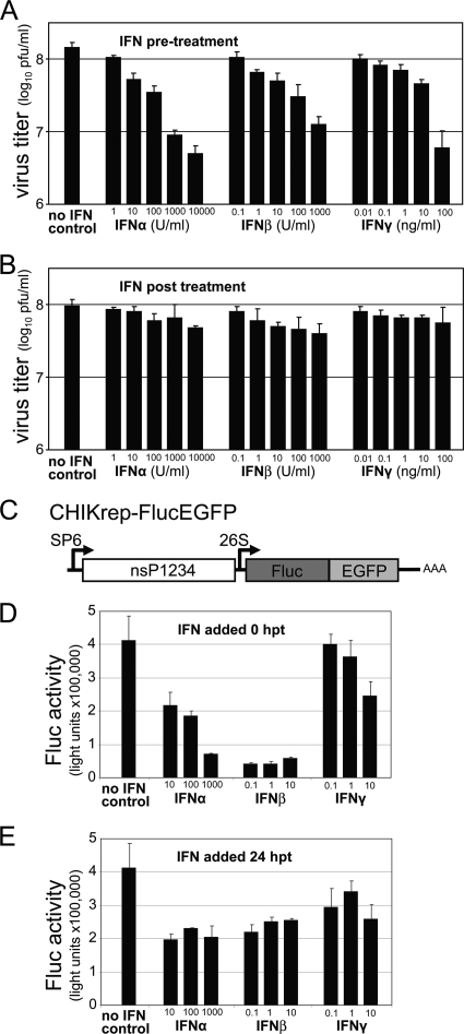
FIG. 1.
Resistance of CHIKV to type I/II IFN treatment. (A and B) Sensitivity of CHIKV infection to IFN treatment. IFNs were added as indicated to CHIKV-infected Vero cells 6 h prior to infection (A) or 4 h p.i. (B). Supernatants were collected 24 h p.i., and virus titers were determined by plaque assays. Error bars represent standard deviations of duplicates. (C) Schematic representation of the CHIKrep-FlucEGFP replicon expressing an Fluc-EGFP fusion protein. (D and E) Sensitivity of the replication of CHIKV replicon RNA to IFN treatment. Different concentrations of IFNs were added to CHIKV replicon-transfected Vero cells in 96-well plates directly posttransfection (0 h p.t.) (D) or 24 h p.t. (E), and Fluc activity was measured 48 h p.t. Concentrations of IFN-α are expressed in international units (IU) per ml, and IFN-β/γ concentrations are expressed in ng per ml. Error bars represent standard deviations.

