FIG. 3.
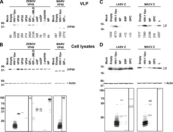
Filoviral GP1,2, but none of the arenavirus-encoded proteins, counteract human BST-2. (A) Human 293 cells stably expressing human BST-2 were cotransfected with ZEBOV HA-VP40 DNA and plasmids encoding ZEBOV VP30-V5, VP35-V5, V5-VP24, GP1,2, GP1,2ΔMLD-V5, sGP-V5, ssGP-V5, Δ-peptide-V5, or NP-V5 or HIV-1 Vpu-V5. Alternatively, cells were transfected with MARV HA-VP40 DNA, together with plasmids encoding MARV GP1,2 or HIV-1 Vpu-V5. Cell lysates and supernatants were treated as in Fig. 1, and HA-tagged VP40 in VLPs were analyzed by Western blotting. Numbers below each lane indicate values obtained with densitometric scanning using the ImageJ program (NIH). (B) Expression of HA-tagged VP40, actin, and other filovirus-encoded proteins in cell lysates was determined by Western blotting. Expression of HIV-1 Vpu (∼16 kDa), ZEBOV VP30 (∼30 kDa), VP35 (∼35 kDa), VP24 (∼24 kDa), GP1,2 ΔMLD (∼75 kDa), sGP (∼50 kDa), ssGP (∼47 kDa), Δ-peptide (∼10 kDa), and NP (∼104 kDa) was detected using anti-V5 antibody. Expression of ZEBOV (∼140 kDa) and MARV (∼170 kDa) GP1,2 was detected using anti-GP antibodies (6D8 and 5D7, respectively). (C) Human 293 cells stably expressing human BST-2 were cotransfected with LASV Z-HA DNA and plasmids encoding LASV NP-V5, SSP-V5, or GPC or HIV-1 Vpu-V5. Alternatively, cells were transfected with MACV Z-HA and MACV NP-V5, GPC, L-FLAG, or HIV-1 Vpu-V5. Cell lysates and supernatants were treated as in Fig. 1, and HA-tagged Z proteins in VLPs were analyzed by Western blotting. Numbers below each lane indicate values obtained with densitometric scanning using the ImageJ program (NIH). (D) Expression of HA-tagged Z, actin, and other arenavirus-encoded proteins in cell lysates was determined by Western blotting. Expression of HIV-1 Vpu (∼16 kDa), arenaviral NP (∼63 kDa), and LASV SSP (∼10 kDa) was detected by using anti-V5 antibody. Expression of MACV L-FLAG (∼200 kDa) was detected using anti-FLAG antibody. Expression of LASV GPC (∼75 kDa) and GP1 (∼42 kDa) was detected with an antibody against GP1 (161-6). Shown is a representative Western blot from three independent experiments.

