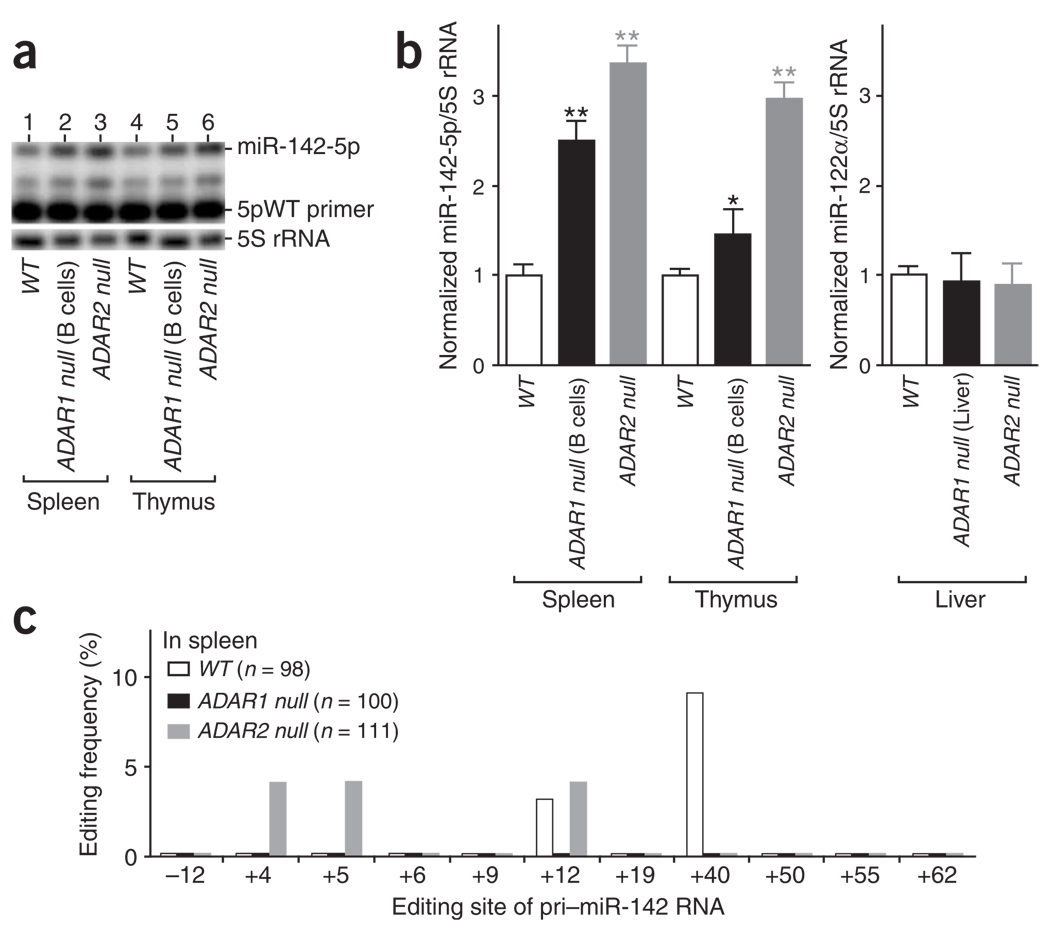
Figure 6.
Increased expression levels of miR-142 RNAs in spleen and thymus of ADAR null mice. (a) The wild-type sequence primer 5pWT was used for quantitation of mature miR-142-5p RNA expression levels as in Figure 2d. Identical results were obtained with a separate extension assay done by using the degenerate sequence primer 5pDG (data not shown). (b) The level of the mature miR-142-5p in wild-type and ADAR null mice. Results from three independent experiments are shown. The increase in the relative miR-142-5p levels in comparison to wild-type mouse tissue was examined statistically by individual unpaired Student’s t-tests. Significant differences are indicated as follows: one asterisk, P < 0.01; two asterisks, P < 0.001. Error bars, s.e.m. (c) The editing pattern (quantitated as in Figure 1c) revealed by sequencing of cDNA clones corresponding to the endogenous pri–miR-142 RNAs edited in vivo in spleens of wild-type, ADAR1 null and ADAR2 null mice.

