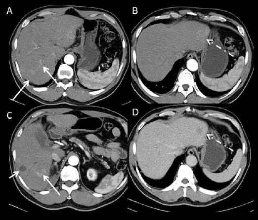
Figure 1.

Dual-phase contrast-enhanced multidetector computed tomography images. (A, B, C): images acquired at the arterial phase. (D): image acquired at the portal venous phase at the same level as B. There is a treated non-enhancing hepatocellular carcinoma in the right hepatic lobe (short arrow on C). There is an enhancing lesion with washout, compatible with hepatocellular carcinoma in the lateral left lobe (dashed arrow). There are also multiple areas of arterial enhancement without washout in the right lobe (long arrows) that are difficult to characterize. Magnetic resonance imaging was performed to further characterize these findings.
