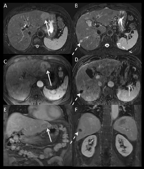
Figure 2.

1.5 Tesla magnetic resonance imaging with an extracellular gadolinium contrast agent (gadopentetate dimeglumine) obtained 6 weeks after computed tomography in the same patient as in Figure 1. (A, B): axial fat suppressed T2-weighted images. (C): axial post-contrast fat suppressed T1-weighted image acquired at the arterial phase. (D): axial T1-weighted subtracted image acquired at the arterial phase at a different level. (E, F): coronal post-contrast fat suppressed T1-weighted images acquired at the portal venous phase. The treated right lobe hepatocellular carcinoma is T2 hyperintense and completely necrotic (dashed arrows on B and D). The left lateral lobe lesion (arrow) measures 4.3 cm and is T2 hyperintense with arterial enhancement and washout compatible with hepatocellular carcinoma. The vague areas of enhancement seen on computed tomography are likely areas of arterioportal shunting, as they are isointense on T2- and post-contrast T1-weighted images acquired at the portal venous and equilibrium phases.
