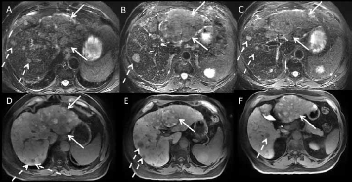
Figure 3.

3 Tesla magnetic resonance imaging with liver-specific gadolinium contrast (gadoxetate disodium) in a patient with cirrhosis and hepatocellular carcinoma. (A, B, C): axial fat suppressed T2-weighted images. (D, E, F): hepatocyte phase T1 post-contrast images (20 min). There is a large infiltrative mass (arrows) of the left hepatic lobe, which is T2 hyperintense with heterogeneous signal on post-contrast hepatocyte phase images, with areas in low and high signal. Areas in high signal presumably represent well-differentiated hepatocellular nodules. In the right lobe, there are innumerable T2 hyperintense nodules (dashed arrows) with decreased gadoxetate disodium uptake on the hepatocyte phase, compatible with multifocal hepatocellular carcinoma. The patient is outside transplant criteria and cannot be resected, and will be treated with radioembolization.
