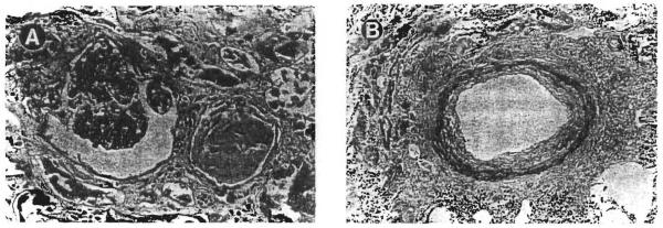
Fig 2.
Case no. 7 showed more pronounced histopathologic changes than those illustrated in Fig 1. (A) The glomeruli showed an accentuation of the lobular pattern and a segmental increase in the mesangial cellularity. The degree of interstitial fibrosis was greater than that observed in case no. 5. (Magnification ×200.) (B) The arteries showed fibrous thickening of the intimal layer. (Magnification ×100.)

