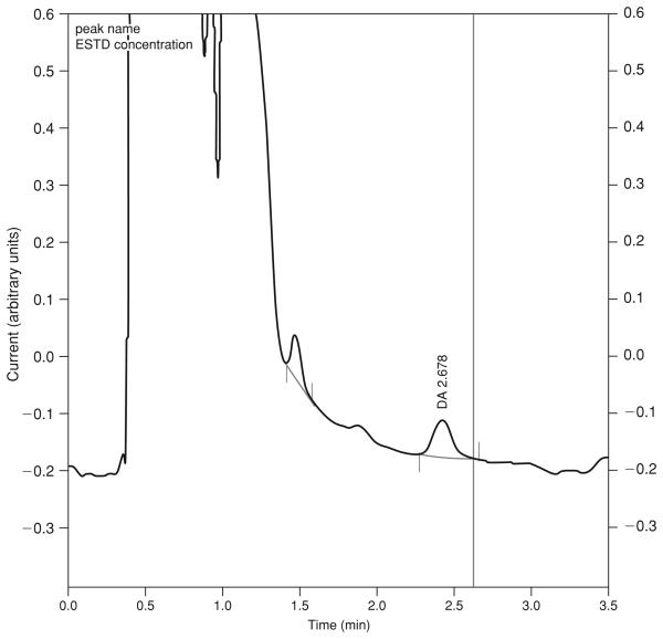
Figure 7.4.3.
Chromatogram for detection of dopamine (DA) in a basal dialysate sample from mouse nucleus accumbens. A 1-mm probe was used. Dialysis flow rate was 0.7 μl/min and 8 μl were injected. Ascorbic acid was included in the aCSF and it elutes as the early, major peak. The number above the DA peak is the dialysate concentration (nM) estimated from the external standard curve. The oxidation potential was set at +700 mV.

